訓練研討會:蔡司晶格光片7顯微鏡
訓練研討會:蔡司晶格光片7顯微鏡
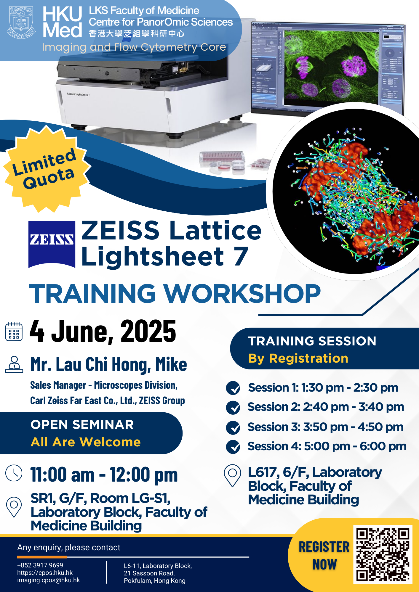
Open Seminar (All are welcome to join!)
揚聲器: Mr. Lau Chi Hong, Mike (Sales Manager – Microscopes Division, Carl Zeiss Far East Co., Ltd., ZEISS Group)
日期: 4 June, 2025
時間: 11:00 am -12:00 pm
地點: SR1, G/F, Room LG-S1, Laboratory Block, Faculty of Medicine Building
Training Session (By registration)
- 註冊者必須參加公開研討會
- CPOS成像和流式細胞儀核心實驗室的成像使用者將優先參加培訓研討會
- Venue: L6-17, 6/F, Laboratory Block, Faculty of Medicine Building
- Maximum 6 seats in a single training session
| Training Session | 日期 | 時間 |
| Session 1 | 4 June, 2025 | 1:30 pm – 2:30 pm |
| Session 2 | 4 June, 2025 | 2:40 pm – 3:40 pm |
| Session 3 | 4 June, 2025 | 3:50 pm – 4:50 pm |
| Session 4 | 4 June, 2025 | 5:00 pm – 6:00 pm |
👉 Reserve Your Spot Now!
註冊連結: https://hkuems1.hku.hk/hkuems/ec_hdetail.aspx?ueid=100304
這 Zeiss Lattice Light Sheet 7 Microscope is a light sheet setup that allows long-term volumetric imaging of subcellular structures in attached or suspended cells, spheroids, or organoids with diameters up to 200 μm.
✅ Long-term live imaging: Integrated incubation and autoimmersion systems ensure experiment stability over extended periods.
✅Reduced photobleaching: Ultra-thin light sheet illumination minimizes photobleaching by 70% compared to widefield microscopy.
✅Deeper penetration: Parallel light sheet illumination reduces scattering, revealing subcellular details within organoids or embryos.
✅High-speed volumetric imaging: Capture full 3D volumes with synchronized light sheet scanning and high-sensitivity cameras.
✅Smart data processing: ZEISS Arivis Pro software automates 3D/4D image reconstruction, GPU-accelerated deconvolution, and AI-based analysis.
The Zeiss Lattice Lightsheet Microscope 7 has been installed in CPOS Image and Flow Cytometry Core.
For more information about instrument specification, please visit 在此
如有任何疑問、預訂或需要協助:
📧 imaging.cpos@hku.hk | 📞 3917 9699


