影像設備
影像與流式細胞術核心實驗室
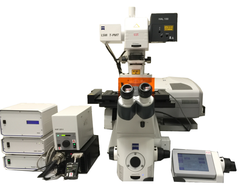 LSM780 is an inverted laser scanning confocal microscope. It is equipped with three solid state lasers (405, 561 and 633 nm), one argon laser with three laser lines (458, 488 and 514 nm), one femtosecond pulsed laser (Coherent Chameleon Vision II, tuning range 680nm-1080nm), two PMT detectors, one 32-channel spectral GaAsP detector and one transmitted light PMT detector. The GaASP detector allows the detection of weak signals and spectral imaging with unmixing (separation of spectrally overlapping fluorophores). The stage-top live cell chamber, the motorized stage, photo-bleaching module enables the multi-dimensional (multi-channel, multi-position, time-lapse) live cell imaging, photon-manipulation and large tissue tile scanning.
Applications:
Standard Operation Protocol – Carl Zeiss LSM780.pdf
Staff-in-charge: Dr. WANG Zhaoyue (Cindy) | 3917 9851 | imaging.cpos@hku.hk
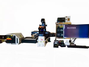
Live-SR Super-resolution/TIRF Microscope is an integrated imaging system equipped with a Live-SR Super-resolution Unit, a Yokogawa CSU-W1 Spinning Disk Unit, and an iLAS3 Ring-TIRF/FRAP/Ablation Unit. The Live-SR is based on an optically demodulated structured illumination technique with online processing. It enables super-resolution (105nm laterally) at high speed with low photo-toxicity. iLAS3 Ring-TIRF/FRAP with 4 laser lines (405nm, 488nm, 561nm, and 639nm) allows the observation of membrane-associated processes like cell adhesion, hormone binding, molecule transport, exocytotic and endocytotic dynamics. A high-power 355nm UV pulsed laser enables ablation for applications like DNA damage and axotomy.
Applications:
Detailed Configuration
Standard Operation Protocol – Live-SR Super Resolution/TIRF Microscope
Staff-in-charge: Mr. WU Kelly | 3917 9445 | imaging.cpos@hku.hk
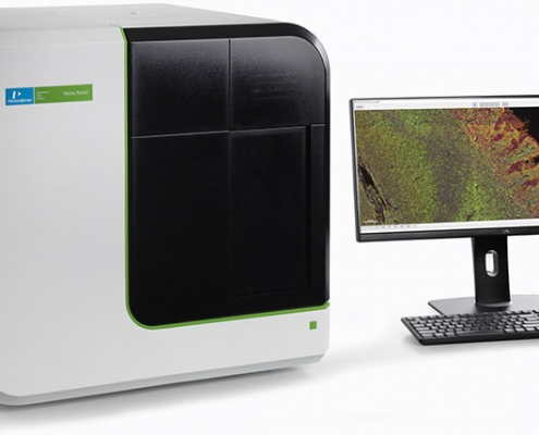 Vectra Polaris integrates both multispectral imaging and automated slide scanning to better visualize and quantify immunolabelled cells in tissue sections and tissue microarrays. With Tyramide signal amplification-based staining of multiple biomarkers (up to 8 overlapping fluorescent colors and DAPI), visualization is achieved by patented Liquid Crystal Tunable filter and unmixing algorithm. This fully automated system provides high speed whole-slide scanning at 10x to 40x in brightfield or fluorescence for up to 80 slides in a run. Supported by the tissue analysis software, quantification and spatial analysis of biomarkers can be accomplished in high throughput via machine learning and batch analysis.
Applications:
Standard Operation Protocol – Perkinelmer Vectra Polaris
Opal staining protocol
Akoya Opal Assay Development_Guide
Staff-in-charge: Dr. WANG Zhaoyue (Cindy) | 3917 9851 | imaging.cpos@hku.hk
The LiTone XL Light-sheet Microscope is a lightsheet setup that applies unique 4-side illumination to achieve sample penetration across the entire sample. The microscope is equipped with 4 solid lasers (405 nm, 488 nm, 561 nm, and 640 nm). With the sCMOS camera and high NA objective, the microscope provides lateral resolution to 500 nm and axial resolution to 1.5 um. The microscope has optics that are adaptable to a wide range of refractive indices, from water to solvent solutions, with an index of refraction ranging from 1.33 to 1.52. With its live cell imaging toolkit and the motorized stage, the microscope enables long-term imaging of live small transparent animal, such as zebrafish. The detachable sample bath and holder set allow for imaging of both soft and hard samples ranging in size from 0.5 mm to 25 mm.
Applications:
Detailed Configuration
Standard Operation Protocol – LiTone XL Light-sheet Microscope
Staff-in-charge: Mr WONG, William | 3910-3523 | imaging.cpos@hku.hk
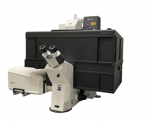
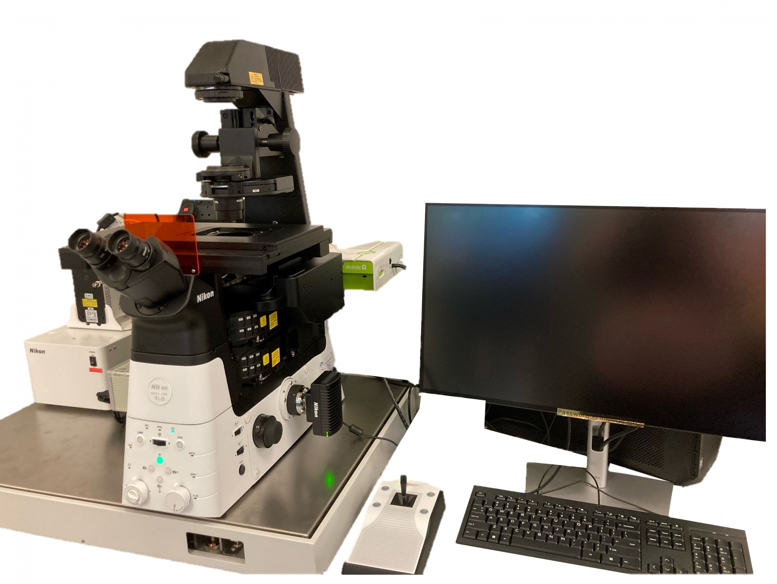
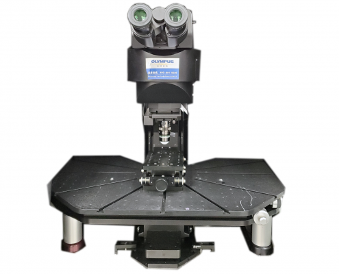 Olympus FVMPE-RS Hybrid Multiphoton System is a multi-photon laser scanning microscope that allows fluorescence imaging deep into specimens at hundreds of micrometers in living cells and tissues. It is equipped with two Coherent Chameleon Vision II laser, tunable from 680nm to 1080nm, four NDD (non-descanned detector) detectors (PMT), upright microscope stand and dual scanners (Galva and Resonant Scanner). This enables simultaneous imaging of maximum four fluorescent channels on live samples. High NA water immersion objectives allow imaging directly into specimens in media. It is dedicated to intravital imaging of living, whole mount, thickly sliced specimens or small animals.
Applications:
Detailed configuration
Standard Operation Protocol – Olympus MP-ERS
Staff-in-charge: Dr. WANG Zhaoyue (Cindy) | 3917 9851 | imaging.cpos@hku.hk
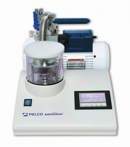
During the process of vitrification, the aqueous sample solution may form a droplet and fail to spread evenly on EM grids. Plasma cleaning is a key step in preparing EM grids for vitrification, especially those with hydrophobic carbon films. The PELCO easiGlow™ Glow Discharge Cleaning System is a highly effective method for cleaning EM grids and turning carbon film surfaces hydrophilic. With a glow discharge treatment, the PELCO easiGlow™ system can modify the surface properties of carbon films, allowing aqueous solutions to spread easily and ensuring even distribution of the sample on the grid. This process is essential for high-quality vitrification and subsequent cryo-EM imaging.
Features
Standard Operation Protocol – PELCO easiGlow
Staff-in-charge: Dr. WANG Zhaoyue (Cindy) | 3917 9851 | imaging.cpos@hku.hk

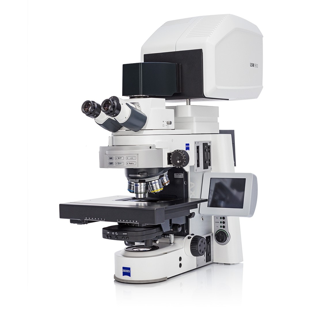
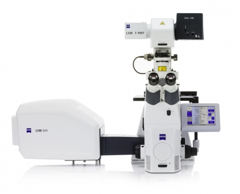 LSM 800 is an inverted confocal setup equipped with 4 lasers (405 nm, 488 nm, 561 nm, 640nm) and Airyscan for sub-diffraction resolution to 140 nm. The microscope has 2 confocal detectors and a highly sensitive GaAsP detector, each of the detector’s wavelengths is tunable per nanometer. The motorized stage, advanced tiling module, photo-bleaching module enables the multi-dimensional (multi-channel, multi-position) imaging, photo-manipulation and big tissue tile scanning. Applications:
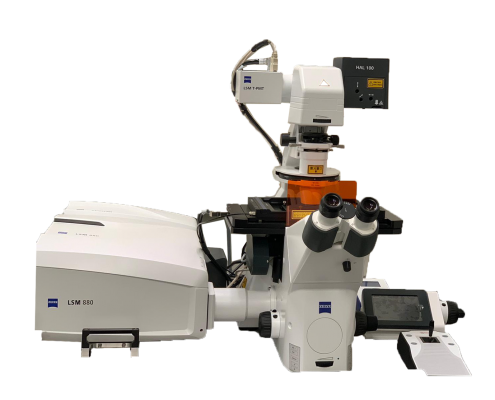
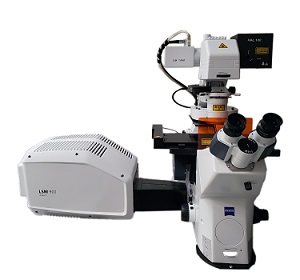
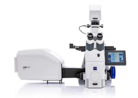
 The Zeiss LSM980 system is an inverted laser scanning confocal microscopes with Airyscan 2 expands the excitation laser spot to allow imaging of eight lines in parallel simultaneously. It enables multiplex modes of SR-8Y, SR-4Y and CO-8Y for high speed imaging without sacrificing sensitivity or resolution. New ZEN software modules, including “Sample Navigator” and “Zen Connect” will simplify the imaging setup procedure. The stage-top live cell chamber, the motorized stage, advanced tiling module, photo-bleaching module enables the multi-dimensional (multi-channel, multi-position, time-lapse) live cell imaging, photo-manipulation and large tissue tile scanning.
 PE UltraVIEW VoX spinning disk confocal is built for rapid and gentle imaging of live cell specimens. Highly sensitive EMCCD camera coupled with Yokogawa CSU-X1 spinning disk unit enables user to image with very low laser power to minimize effect of phototoxicity during long-term live cell experiments. Piezo z-stage is included in the setup for capturing volume up to 400 µm in z-axis with exceptional speed.
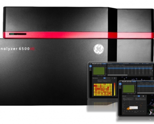 IN Cell Analyzer 6500HS is a laser-based line scanning high-content imaging system. High speed filter based widefield, EDGE confocal imaging modes with a sensitive PCO Edge 4.2 sCMOS camera. User could choose between fixed cell imaging, live cell incubation and hypoxic incubation ([Oxygen] adjustable 0.1% -20%). Combined with new IN Carta software, for object quantification streamlines workflow for high content analysis feature extraction.
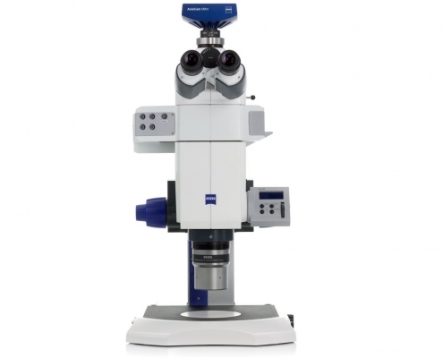 AxioZoom.V16 is a multi-zoom stereoscope with fluorescent illumination. High N.A. optics enable visualization of specimen ranging from large field-of-view to single cell observation. Flexible design allows illumination options of brightfield, dark field, oblique ring illumination, reflective fluorescence for DAPI / GFP / RFP.
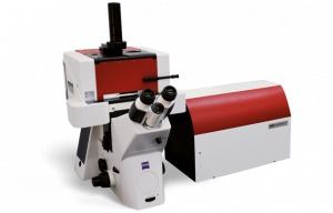